
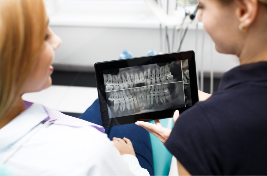
A 3D dental scan can change the treatment plan for a difficult implant case by showing the height and width of the jawbone, where nerves run, and how much room is available. The implant can then be repositioned, resized, or delayed for a bone graft.
A 3D implant scan removes guesswork from complex cases and makes the result more predictable for the patient. A consultation explains what the images show, how the plan may change, and which option suits the mouth.
How a 3D Scan Fits Into a Difficult Implant Case
Standard X-rays are flat, so they show height but not thickness. A cone beam CT, or CBCT, builds a three-dimensional picture of the jaw that can be turned and measured from any angle.
For a difficult implant case, that view often changes the plan. Bone that looked sufficient on a flat image may turn out thin, or a spot that looked tight may have more room than expected.
Which Details a CBCT Scan Shows About Bone and Nerves
CBCT implant planning starts with jawbone anatomy, since the scan measures how tall, wide, and dense the bone is at the implant site. It also shows implant nerve location, so the implant can sit a safe distance from the nerve in the lower jaw.
Why Limited Bone Changes the Plan
When a bone evaluation for a dental implant shows thin or short bone, the plan often shifts. A bone graft may be recommended to build the area before placement, or a different implant size may be chosen.
Seeing this before surgery lets patients discuss the extra step and its timing in advance, rather than discovering the problem on the day.
How Digital Planning Turns Images Into a Surgical Roadmap
Digital implant dentistry combines the scan with planning software. The implant is positioned on screen first, so its depth, direction, and spacing from neighboring teeth are worked out before anything is placed.
Complex implant planning benefits most from this step. Several implants, a full arch, or a site with limited space all become easier to organize when the whole layout can be reviewed in 3D.
Which Complex Cases Benefit Most From a 3D Scan
A 3D scan is especially helpful for patients who have been missing teeth for years, those with thin bone, and anyone replacing teeth near the sinus or a nerve. It also helps in full-arch cases.
Simple single-tooth cases may not always need the same level of detail. The dentist recommends imaging based on the patient’s history and the site being treated.
How Robotic Guidance Builds on 3D Imaging
Some practices pair 3D imaging with robotic-assisted guidance during placement. The scan provides the plan, and the guidance helps the implant follow it during surgery.
This pairing supports a more predictable result because the placement reflects the planning done beforehand.
How Follow-Up Care Protects a Planned Implant
After placement, regular visits let the dentist check healing and the bite. Restorative dentistry then completes the implant with a denture, custom crown or bridge.
Preventive dentistry and cleanings keep the gums healthy, which supports long term care of the implant. Follow up care also lets small changes be caught early.
3D Implant Planning at Dental Excellence of Hatfield
At Dental Excellence of Hatfield in Hatfield, PA, we offer dental implants planned with a Carestream CBCT 3D scanner. We also use the Yomi robotic system, which combines detailed 3D imaging with robotic guidance for implant planning and placement.
Our office at 461 S Main St welcomes patients with difficult or long-standing tooth loss, and sedation options are available when appropriate. Patients are welcome to ask us how a 3D scan could shape their own implant plan.
Implant Consultations in Hatfield, Colmar and Lansdale
Adults in Hatfield, Colmar and Lansdale who have been told their implant case is complicated are welcome to book a consultation with our team to see how a 3D scan could clarify the plan and the next steps.
Frequently Asked Questions
How does imaging help evaluate the sinus area before an upper implant?
A 3D scan shows how close the sinus floor is to the planned site and how much bone lies beneath it. That helps decide whether the implant fits or the area needs added support first.
How does digital implant planning help determine the angle of the implant?
The software lets the implant be tilted and positioned on screen against the bone and neighboring teeth. This confirms a safe angle that supports the future crown before any surgery begins.
How is radiation exposure kept as low as practical during 3D imaging?
Scans are taken only when needed, focused on the area being treated and set to the lowest settings that still give clear images. Protective gear and short scan times also reduce exposure.
Can the final crown position be considered before the implant is surgically placed?
Yes. Planning begins with where the finished tooth should sit, and the implant is positioned to support it. This keeps the crown, bite and smile in mind from the start.